
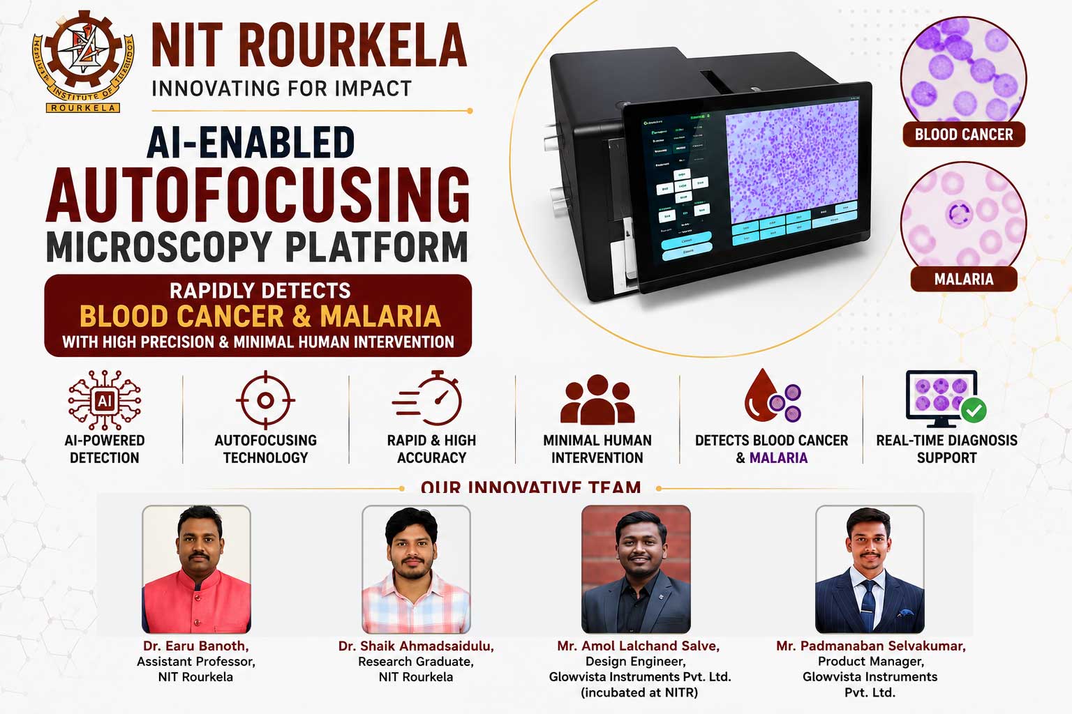
NIT Rourkela researchers have created an AI-enabled autofocusing microscopy platform that rapidly detects blood cancer and malaria with high precision and minimal human intervention.
AI-Enabled Autofocus Transforms Microscopic Imaging
Researchers at the National Institute of Technology (NIT), Rourkela, have developed an artificial intelligence (AI)-enabled autofocusing technology that significantly improves microscopic imaging for biomedical diagnostics. The system has already demonstrated promising lab-scale results in detecting acute lymphoblastic leukaemia (blood cancer) and malaria, marking a major step toward affordable next-generation healthcare diagnostics in India.
Developed in collaboration with Glowvista Instruments Private Limited, a startup incubated at NIT Rourkela’s Incubator Centre (FTBI), the new technology produces rapid, accurate, and repeatable results with minimal human intervention. The research team has secured a patent for the technology.
Limitations of Conventional Microscopy Systems
Microscopy technology plays a crucial role in healthcare diagnostics by enabling doctors and researchers to study cells, tissues, microorganisms, and other biological structures invisible to the naked eye. It identifies cancers, infectious diseases such as malaria and tuberculosis, pathology-related disorders, and supports drug development and point-of-care diagnostics.
However, conventional microscopy systems largely depend on manual focusing, making them time-consuming and vulnerable to human error. These limitations often result in inconsistent imaging, inaccurate diagnoses, and treatment delays – potentially fatal when dealing with complex biological samples or emergency diagnostic requirements.
Optofluidic Digital Microscopy with AI Feedback
Earu Banoth, assistant professor at NIT Rourkela and founder-director (non-executive) of Glowvista Instruments Pvt Ltd, said the team developed an optofluidic digital microscopy platform to address longstanding limitations in conventional microscopy systems.
“The optofluidic digital microscopy platform combines deep-learning algorithms with optical imaging systems and automated motion control. The intelligent system continuously analyses microscopic images in real time and automatically adjusts focus through an AI-driven feedback mechanism, improving imaging precision and efficiency,” he said.
Built at a cost of around ₹1.2 lakh, the system demonstrated promising results during laboratory testing. Besides detecting acute lymphoblastic leukaemia and malaria, it successfully carried out complete blood cell counts through five-class and seven-class blood cell categorisation.
Handheld System Matching Imported Technologies
Explaining the broader objective, Banoth said the team is working toward developing a simple handheld system that performs at par with expensive imported automated microscopy technologies while offering precise diagnostic information. “The technology is also being designed for wider biomedical applications beyond the capabilities of conventional flow cytometers and imaging flow cytometers,” he said.
The developed system incorporates several advanced features, including AI-powered intelligent autofocus with real-time image processing, automated motion control for precise focus adjustment, enhanced imaging of complex biological samples, cloud-enabled learning for continuous improvement, and user-friendly operation with improved repeatability and efficiency.
Broad Applications Across Healthcare and Life Sciences
According to the researchers, the technology has potential applications across healthcare and life sciences fields, including biomedical diagnostics, digital pathology, AI-assisted microscopy, automated imaging, point-of-care healthcare devices, biofluid monitoring, smart laboratory automation, and portable remote diagnostic systems.
“We are now working on generating larger ground-truth datasets and scaling up the system for field deployment in diagnostic centres and research laboratories. Feedback from field trials will be used to refine the technology and secure approvals for commercial deployment,” said Banoth.
The researchers are also seeking additional support from research and startup ecosystems to accelerate the development of a market-ready product. Other team members include Shaik Ahmadsaidulu, research graduate; Amol Lalchand Salve, design engineer; and Padmanaban Selvakumar, product manager, from Glowvista Instruments Pvt Ltd.
The research received funding support from the Anusandhan National Research Foundation (ANRF), Department of Science and Technology (DST), and Department of Biotechnology (DBT) under the Government of India.
#NITRourkela #AIPoweredMicroscopy #AutofocusTechnology #BloodCancerDetection #MalariaDiagnosis #GlowvistaInstruments #DigitalPathology #PointOfCareDiagnostics #AIHealthcare #BiomedicalDiagnostics
Disclaimer
The information in this article is based on available public sources and official statements as of the time of publication. While we aim for accuracy, we do not guarantee completeness or correctness. We advise readers to verify key details from official sources before making any decisions. The website (iitiimsamvaad.com) is not liable for any loss or damage arising from the use of this content. The authors are also not responsible for any such loss or damage.